
Share this page
Spinal Deformity
Adolescent Idiopathic Scoliosis
A three-dimensional curvature of the spine that develops during the adolescent growth spurt — usually mild, painless, and entirely compatible with a full, active life.
For information only — not medical advice. The content on this page is patient education to help you understand a condition or treatment. It is not a substitute for personalised assessment by a qualified clinician. Always consult your doctor or specialist about your own symptoms, diagnosis, and treatment decisions.
Patient leaflets
Download and take away
Print-friendly guides written for patients and families — save them, share with your GP, or bring them to your consultation.
Please read first
A note before you start
Scoliosis is one of the most talked-about spinal conditions, and also one of the most misunderstood. For most young people who have it, it is mild, painless, and entirely compatible with a full, active life. This page explains what adolescent idiopathic scoliosis actually is, why it happens, how it is assessed, and what can be done about it — written for both teenagers and the parents supporting them.
This information is for general education and does not replace the advice of your own clinical team. Every scoliosis case is different, and any plan is tailored to the individual. You should seek a medical review if you notice a rapid change in the shape of the back, pain that is persistent or interferes with sleep, or any neurological symptoms such as weakness or numbness in the legs.
What is scoliosis?
Scoliosis is a sideways curvature of the spine, but that simple description undersells it. It is really a three-dimensional change in the shape and position of the spine and trunk — a curve that is also accompanied by a subtle twist. A medical diagnosis is confirmed when a standing X-ray shows a side-to-side curve measuring 10 degrees or more (the "Cobb angle"), together with rotation of the individual vertebrae.
The most common form by far is adolescent idiopathic scoliosis, usually abbreviated to AIS. "Adolescent" reflects the age at which it appears — typically between about 10 and 18 years, coinciding with the rapid growth spurt of puberty. "Idiopathic" simply means the cause is unknown; it tends to appear in children who are otherwise completely healthy. Idiopathic cases account for roughly 80% of all scoliosis.
It is worth knowing that not every curve is the same kind. Doctors distinguish structural scoliosis — a fixed, genuine change in the anatomy of the spine, which includes that characteristic twist — from functional scoliosis, a temporary curve caused by something outside the spine, such as one leg being slightly shorter than the other. A functional curve straightens out when the underlying cause is addressed or when the person lies down; a structural curve does not. Adolescent idiopathic scoliosis is structural.
Who it affects
Scoliosis affects roughly 2 to 3 percent of the general population, which makes it far more common than many people realise. It is grouped by the age at which it first appears — infantile (birth to three), juvenile (four to ten), adolescent (eleven to eighteen) and adult — with the adolescent form being by far the most common.
For mild curves, boys and girls are affected at similar rates. The striking difference is in how curves behave over time: girls are significantly more likely to develop curves that progress and need treatment. For small curves the ratio of girls to boys is roughly even, but among severe curves girls outnumber boys several times over.
Scoliosis also tends to run in families. The genetic contribution is estimated at around 38 percent, and it behaves as a complex, polygenic trait — there is no single "scoliosis gene," but rather many genetic markers that together influence how bone and cartilage develop. If a parent has scoliosis the risk to their child is modestly higher, and a brother or sister of an affected child has a meaningfully raised chance of having a curve too.
A few associations are worth mentioning, mainly so they aren't misread as causes. AIS is seen a little more often in adolescents with a lower body weight, and in certain sports that select for slim, flexible, fast-growing young people — rhythmic gymnastics, ballet, swimming. These are associations, not causes; the sport does not create the curve.
What's happening in the spine
The key to understanding scoliosis is that it is not a simple lean to one side. It is a change in three planes at once.
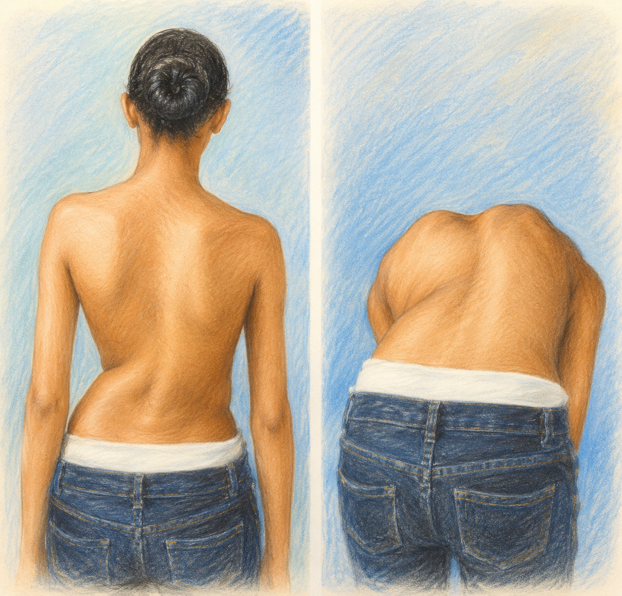
The first and most visible is the side-to-side curve — the lateral deviation that gives scoliosis its name. The second is rotation: as the spine curves, the vertebrae also twist along their axis. The front of each bone rotates toward the outside of the curve while the bony bumps you can feel down your back rotate toward the inside. Because the ribs are attached to these vertebrae, they twist too, and on one side they are pushed backward — producing the characteristic "rib hump" that becomes visible when a person bends forward. The third change is front-to-back: the natural rounded curve of the upper back often flattens, and in some cases reverses slightly, a pattern sometimes called "flat back."
Underneath these visible changes, the anatomy itself remodels. Where the curve is tightest, the vertebrae are under more pressure on the inside and grow more slowly there, while growing faster on the outside — so the bones gradually become wedge-shaped. The front of the spine often grows faster than the back, which contributes to the flattening of the upper back. Over time the muscles and ligaments on the inside of the curve shorten and tighten, which helps to "set" the deformity. A helpful way to picture the whole process is a column that, growing slightly faster on one side than the other, has no option but to curve and twist as it searches for room to grow.
Why it happens — and what doesn't
The honest answer to "why" is that we do not fully know — that is exactly what "idiopathic" means. When the curve appears in an otherwise healthy child, with no underlying bone malformation or nerve disorder to explain it, that is the diagnosis.
What researchers do believe is that scoliosis is multifactorial — the result of several biological factors acting together rather than a single trigger. Leading lines of study include subtle disproportions in how the front and back of the growing spine develop, hormonal signals during puberty (such as sensitivity to leptin or imbalances in melatonin) that influence how bone matures, and small differences in how the brain coordinates balance and posture during a phase of very rapid growth. Layered on top of all this is the familial, genetic tendency described above.
Just as important is what scoliosis is not caused by, because families often carry unnecessary guilt about this. Idiopathic scoliosis is not a lifestyle condition, and nothing in a child's daily habits brings it on. Poor posture does not cause it — slouching can make a back look uneven, but the curve is an internal change in the spine, not the result of sitting badly. Heavy school bags can cause a temporary lean but not this permanent, three-dimensional curve. Sport does not cause it or worsen it. And there is no evidence that the type of mattress or bedding plays any role at all. You did not cause this, and your child did not cause it.
Spotting the signs
Scoliosis is usually painless, which is part of why it can go unnoticed — there is often no symptom to prompt a child to mention it. Instead, it tends to be spotted by its appearance. Common signs include one shoulder sitting higher than the other or an uneven waistline; a "hump" on one side of the back that becomes obvious when the person leans forward (the basis of the Adam's forward bend test); and clothes that hang unevenly, or an arm that brushes against one hip but not the other.
Early detection matters. Most mild curves never cause long-term problems, but spotting a curve while a child is still growing means it can be watched and, if needed, treated before it has the chance to progress.
How it's assessed and measured
Assessment begins with a careful clinical examination, looking for asymmetry in the shoulders, shoulder blades and waist. The most familiar test is the Adam's forward bend test: bending forward at the waist makes any rotation in the spine more obvious, as the rib hump on one side comes into view. A small handheld device called a scoliometer can be rested on the back during this bend to measure the angle of trunk rotation in degrees.
The definitive measurement comes from a standing X-ray, on which the specialist measures the Cobb angle — the size of the side-to-side curve. A curve of 10 degrees or more confirms the diagnosis. Because scoliosis is so closely tied to growth, the specialist also judges how much growing is left to do, using markers of skeletal maturity such as the Risser sign (seen on a pelvis X-ray) or the Sanders staging (based on the small bones of the hand). Knowing where a child sits on their growth curve is central to predicting what a scoliotic curve is likely to do next.
In most straightforward cases, this is all that is needed. An MRI is reserved for situations where there are atypical features — an unusual curve pattern, any neurological signs, significant pain, or a very young child — or as part of the work-up before surgery, to make sure nothing else needs to be addressed first.
Will it change over time?
Whether a curve progresses comes down mainly to two things: how large it is when first found, and how much growing the child has left to do. The risk is highest during the pubertal growth spurt, when the spine is changing fastest. Once growth is complete, the chance of significant further change drops sharply for most people.
This is where reassurance is warranted. Many small curves — those under about 20 degrees — stay stable and never need any treatment at all. Across the published evidence, somewhere between a quarter and three-quarters of curves do not progress. Monitoring exists precisely so that the minority that do start to change can be picked up early, while the majority that stay put can simply be left alone.
Monitoring (watchful waiting)
Management is guided by the size of the curve and the amount of growth remaining. There are three broad approaches, and most young people only ever need the first.
For smaller curves in a still-growing child — typically under about 20 to 25 degrees — the right course is often to do nothing actively, but to keep an eye on things. This means a clinical check and an occasional X-ray every 6 to 12 months to see whether the curve is changing. For most, it never does.
Bracing
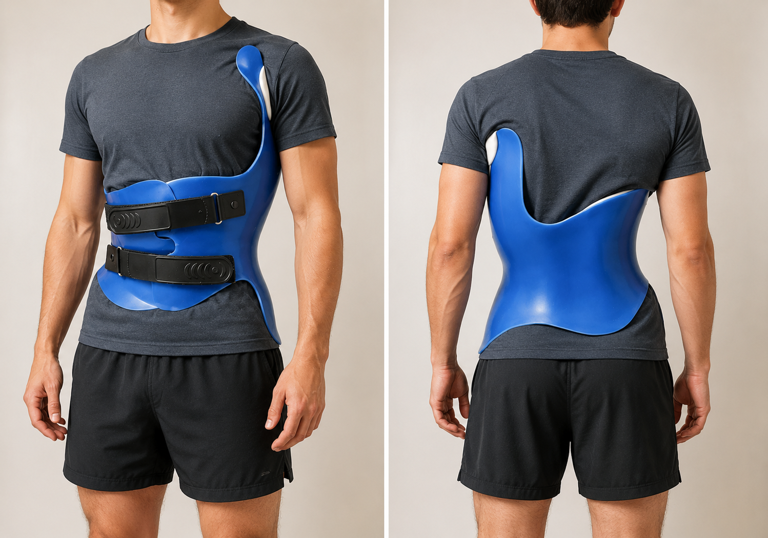
When a curve is in the middle range — roughly 25 to 40 degrees — in a child who is still growing, a brace may be recommended. It is important to understand what a brace is for: its goal is to hold the curve and stop it reaching the size where surgery would be considered, not to straighten or permanently reverse it. A brace works by applying gentle, well-directed pressure to the trunk, and by taking advantage of the fact that a growing bone responds to load — easing pressure on the inside of the curve encourages more even growth, steering the spine as it matures.
The single biggest factor in whether a brace works is how many hours a day it is worn. The landmark BrAIST trial found that bracing roughly increased the chance of avoiding the surgical threshold — about 72 percent of braced children reached maturity without their curve exceeding 50 degrees, compared with about 48 percent of those simply observed — and the benefit climbs steeply with wear time, reaching around 90 percent in those who wore the brace more than about 13 hours a day.
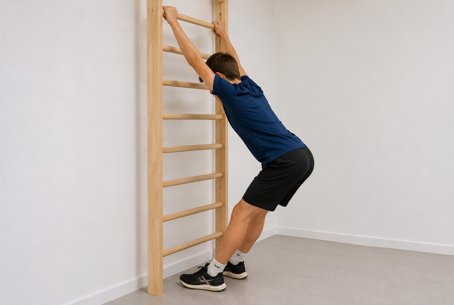
Bracing is frequently paired with scoliosis-specific physiotherapy (approaches such as Schroth or SEAS), tailored exercises that teach a young person to actively correct and stabilise their own posture.
Scoliosis-specific physiotherapy
Programmes such as Schroth and SEAS teach young people exercises specifically designed for the three-dimensional shape of their own curve. The aim is to lengthen the spine, derotate the trunk and rebuild the muscular control needed to hold a corrected posture in daily life. These exercises are usually taught by a certified physiotherapist over a series of sessions and then continued as a home programme — they work best when practised consistently and are often used alongside bracing.
Surgery
Corrective surgery is generally only discussed for larger curves — those exceeding around 45 to 50 degrees, or curves progressing despite other measures. The aim is to correct the deformity as far as is safe, halt its progression and balance the trunk over the pelvis, using modern instrumentation to hold the spine in its new position while it heals.
Recovery is quicker than many families expect: most young people are walking within a day or two, back at school within a few weeks, and returning to sport within a matter of months. In experienced hands these are very safe operations, with careful monitoring used throughout to protect the nerves.
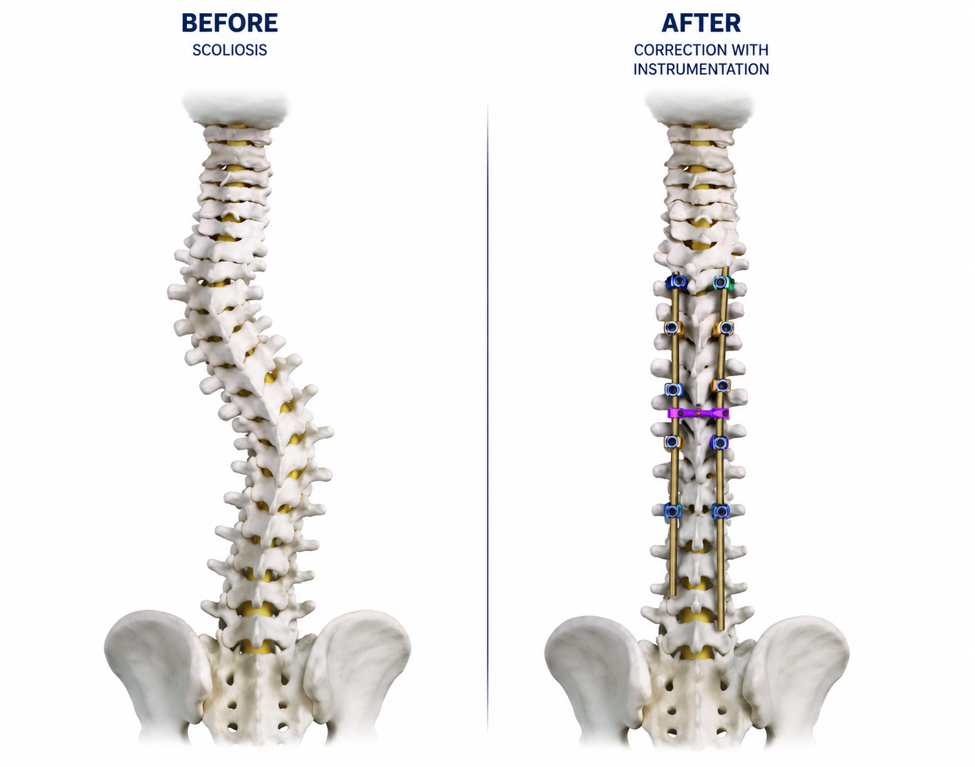
What modern correction looks like
Below are paired standing X-rays of the same young person before and after posterior spinal fusion. The pre-operative image shows the structural curve; the post-operative image shows the corrected spine with the screws and rods that hold the correction while the bone fuses solidly over the following months.
Scoliosis Surgery — Procedure Information
These notes explain the specific operations we may use to correct scoliosis, written to support the discussion we have in clinic. They cover four approaches: single-stage posterior spinal fusion; two-stage anterior thoracic release followed by posterior spinal fusion; single-stage anterior (thoracolumbar) instrumented fusion; and two-stage anterior (thoracolumbar) fusion followed by posterior spinal fusion.
A point that applies to all of them: scoliosis surgery is undertaken to control the deformity, prevent long-term progression and optimise the alignment and balance of the spine — it is not an operation for back pain. While some people do find their back feels more comfortable once the spine is straighter and better balanced, pain relief cannot be the primary reason to proceed.
For context, once an idiopathic curve passes about 50 degrees in a skeletally mature patient, the long-term natural history is a well-documented tendency to keep progressing into adult life — on the order of about 1 degree per year — which over time can lead to increasing deformity, trunk shift, rib prominence and cosmetic asymmetry. Non-operative measures (physiotherapy, scoliosis-specific exercises, activity modification and simple pain relief) may help with comfort and function but have not been shown to prevent progression once a curve is this large, and bracing is generally not effective in skeletally mature patients or in larger curves above about 45–50 degrees. This is why surgery is considered for curves of this size, and which operation is right depends on the size, stiffness and pattern of the curve — a decision we make together.
1. Single-stage posterior spinal fusion (PSF)
When this is used. Posterior spinal fusion is the standard operation for idiopathic curves of surgical size that can be adequately corrected from the back in a single procedure.
What the operation involves. We straighten the spine from the back in one operation. You are positioned on your front, and we make an incision along the midline to expose the bones of the spine. Screws are placed into the vertebrae and connected with rods, so that the curve can be carefully straightened and balanced.
We then place bone graft along the spine to make the treated vertebrae fuse into one solid unit. This comes partly from your own bone, which we gently remove from the small joints at the back of the spine (the facet joints) during the operation, mixed with a processed demineralised bone-matrix graft material that stimulates healing and acts as a scaffold for your own bone to grow across the fused levels. The muscles and skin are then closed, and the spine strengthens over the following months as the fusion heals.
The aim. To correct the alignment of the spine as safely as possible, restore overall balance, reduce rib prominence and cosmetic asymmetry where appropriate, and fuse the involved vertebrae so that the curve does not continue to worsen over the rest of your life.
Risks, and how we minimise them:
• Neurological injury to the spinal cord or nerve roots — which can cause weakness, numbness or, in the most severe cases, paralysis. This is rare: new neurological deficits occur in well under 1% of idiopathic cases, and permanent spinal cord injury is rarer still. Minimised by multimodal neuromonitoring throughout the operation and meticulous surgical technique.
• Infection — minimised by antibiotics around the time of surgery and strict sterile technique (reported infection rates are around 1–2%).
• Bleeding / blood loss — minimised by cell salvage (recycling your own blood) and careful blood management.
• Implant-related problems — such as screw malposition or rod breakage. Minimised by careful, monitored screw placement and modern, rigid fixation.
• Failure of the bones to fuse (pseudarthrosis) — which can require further surgery. Minimised by thorough bone grafting (your own bone plus a demineralised bone-matrix scaffold) and rigid fixation.
• Residual deformity or shoulder imbalance — minimised by careful surgical planning and balancing of the correction.
• Gradual wear of the unfused levels above and below the fusion over many years. Minimised by fusing as few levels as safely possible.
• Temporary effects on breathing after a thoracic fusion. Minimised by early breathing physiotherapy on the ward.
• General risks of any major operation — bleeding, scarring, wound-healing problems, blood clots and anaesthetic risks. Minimised by standard surgical and anaesthetic precautions and early mobilisation with the physiotherapy team.
Large multicentre series in idiopathic scoliosis report overall complication rates of around 6%; more contemporary series report major complication rates of 1–2%, neurological events below 1%, and infection rates of 1–2%.
2. Two-stage anterior thoracic release followed by posterior spinal fusion
When this is used. This planned two-stage approach is used for a thoracic curve that is both large and structurally stiff, with significant rotational deformity and rib prominence. In curves of this size, posterior correction alone may not achieve an adequate result, because the spine is too rigid to straighten fully from the back.
Stage one — anterior thoracic release. We begin by approaching the front of the spine through the side of the chest, and loosen the tight discs on the convex side of the curve. This makes the spine more flexible, so that the second stage can achieve a balanced and controlled correction, and it reduces the forces required to correct a stiff thoracic curve. Where appropriate, we also address the rib prominence with a costoplasty (re-contouring the prominent ribs).
Stage two — posterior spinal fusion. A few days later, we straighten the spine from the back. You are positioned on your front, and we make a midline incision to expose the spine. Screws are placed into the vertebrae and connected with rods so the curve can be carefully straightened and balanced, and we place bone graft (your own bone from the facet joints, mixed with a demineralised bone-matrix scaffold) so the spine fuses solidly over the following months.
The aim. To achieve a safer, more controlled correction of a large, stiff thoracic curve than would be possible in a single operation — restoring balance, reducing rib prominence and cosmetic asymmetry where appropriate, and fusing the involved vertebrae so the curve does not continue to progress.
Risks of the first stage (anterior thoracic release, through the chest):
• Chest and breathing problems — temporary changes in lung function, collapse of the lung on the side of the operation, and a small risk of pneumonia. Minimised by careful technique, a chest drain where needed, and early breathing physiotherapy.
• Injury to structures within the chest — including the lung, major blood vessels and the oesophagus. This is rare. Minimised by meticulous technique and specialist anaesthetic and surgical support.
• Temporary numb patches on the chest wall — due to rib dissection.
• General risks of any major operation — bleeding, infection, scarring, wound-healing problems, blood clots and anaesthetic risks. Minimised by standard precautions, antibiotics and early mobilisation.
Risks of the second stage (posterior spinal fusion):
• Neurological injury to the spinal cord or nerve roots — weakness, numbness or, rarely, paralysis (new deficits in well under 1% of cases; permanent cord injury rarer still). Minimised by multimodal neuromonitoring and meticulous technique.
• Infection — minimised by antibiotics and sterile technique.
• Implant-related problems (screw malposition, rod breakage) — minimised by careful, monitored placement and rigid fixation.
• Failure of fusion (pseudarthrosis) requiring further surgery — minimised by thorough bone grafting and rigid fixation.
• Residual deformity or shoulder imbalance — minimised by careful planning and balancing of the correction.
• Gradual wear of the unfused levels above and below over many years — minimised by fusing as few levels as safely possible.
About the two-stage strategy: because this involves two operations, the overall risk is slightly higher than for a single posterior fusion. However, in very large or stiff thoracic curves the staged approach offers a safer and more controlled correction, reduces the forces placed on the spinal cord, and lowers the likelihood of a significant residual deformity compared with attempting full correction in one operation.
3. Single-stage anterior (thoracolumbar) instrumented fusion
When this is used. This approach is used when the thoracolumbar component of the curve is large, rotated and structurally stiff, and where we want to preserve as many mobile lumbar segments as possible. A posterior-only operation would often need to fuse lower into the lumbar spine than we would wish; correcting from the front lets us avoid that.
What the operation involves. We perform the correction in a single stage from the front. Through an incision on the side of the abdomen or lower chest, we gently move the abdominal contents and, in some cases, the diaphragm, aside to reach the front of the spine. The discs on the convex side of the curve are removed, and the segment is corrected and stabilised from the front with screws and a rod. At the lower lumbar levels, where extra support is helpful, we may insert an interbody cage from the front. This allows us to correct the deformity at the apex of the curve without extending the fusion further down the lumbar spine, preserving lower-back movement.
The aim. To correct the curve effectively while preserving as much lumbar mobility as possible, with the understanding that a subsequent posterior fusion is occasionally required (see risks).
Risks, and how we minimise them:
• Chest and breathing problems — because the approach enters the chest/abdominal cavity, there is a risk of a pneumothorax (air around the lung) and collapse of the lung on the side of the operation, which is why a chest drain is routinely placed at the end of surgery; the lung is then fully re-expanded. Temporary changes in breathing and a small pleural effusion (fluid around the lung) can also occur. Minimised by careful technique, the routine chest drain, and early breathing physiotherapy.
• Vascular or visceral injury — including injury to major blood vessels or abdominal structures. This is rare in modern specialist centres. Minimised by meticulous technique and specialist support, with repair at the time if it occurs.
• Dural tear and CSF (spinal fluid) leak — uncommon in anterior surgery; repaired at the time if it occurs.
• Insufficient correction or imbalance once upright — there is a recognised possibility that correction from the front alone may not fully balance the spine, in which case a posterior fusion may be needed at a later stage. This is uncommon but recognised.
• Implant-related problems (screw malposition, rod breakage) — minimised by careful placement and rigid fixation.
• Failure of fusion (pseudarthrosis) requiring revision — minimised by thorough grafting and rigid fixation.
• Gradual wear of the levels above and below the fusion over many years — minimised by fusing as few levels as safely possible (the very reason for choosing this motion-preserving approach).
• General risks of any major operation — bleeding, infection, scarring, wound-healing problems and anaesthetic risks. Minimised by standard precautions, antibiotics and early mobilisation.
The advantage of this approach is that it corrects the curve while preserving lumbar mobility — with the understanding that a posterior fusion may occasionally be required afterwards.
4. Two-stage anterior (thoracolumbar) fusion followed by posterior spinal fusion
When this is used. This planned two-stage approach is used when the curve has structural involvement extending into the lumbar spine — where the lumbar component is rotated, stiff and structurally significant. A posterior-only approach would likely require fusing lower into the lumbar spine than we would wish, so to preserve as many mobile lumbar segments as possible we correct the lumbar curve from the front first, then complete and stabilise the correction from the back.
Stage one — anterior thoracolumbar fusion. Through an incision on the side of the abdomen or lower chest, we move the abdominal contents and, where needed, mobilise the diaphragm to reach the front of the spine. The discs on the convex side of the thoracolumbar curve are removed, the segment is corrected and structural support is placed; to address the alignment at the base of the curve, we may insert an interbody cage from the front. This corrects the lumbar component effectively and avoids fusing unnecessarily low down the lumbar spine.
Stage two — posterior spinal fusion. A few days later, we achieve the remainder of the correction from the back. You are positioned on your front, and a midline incision exposes the spine. Screws are placed into the vertebrae and connected with rods to straighten and balance both the thoracic and lumbar portions of the curve, and bone graft (your own bone from the facet joints, mixed with a demineralised bone-matrix scaffold) is placed so the spine fuses solidly. The aim is a balanced correction while preserving motion segments lower in the lumbar spine.
The aim. To correct a curve with a significant, stiff lumbar component while preserving as much lower-lumbar movement as possible, achieving a balanced, lasting correction across two controlled stages.
Risks of the first stage (anterior thoracolumbar fusion):
• Abdominal-approach effects — because we work near the abdominal contents and diaphragm, there can be temporary slowing of the bowel, and numbness or weakness of the abdominal wall. Minimised by careful technique; these effects are usually temporary.
• Vascular or visceral injury — including injury to major blood vessels or abdominal organs. Rare. Minimised by meticulous technique and specialist support, with repair at the time if needed.
• Chest and breathing problems — if the diaphragm is mobilised, there may be temporary changes in breathing or a small pleural effusion; there is a risk of lung collapse or pneumothorax, so a chest drain is placed for a few days to help the lung re-expand. Minimised by the routine chest drain and early breathing physiotherapy.
• General risks of any major operation — bleeding, infection, scarring, wound-healing problems, blood clots and anaesthetic risks. Minimised by standard precautions, antibiotics and early mobilisation.
Risks of the second stage (posterior spinal fusion):
• Neurological injury to the spinal cord or nerve roots — weakness, numbness or, rarely, paralysis (new deficits in well under 1% of cases; permanent cord injury rarer still). Minimised by multimodal neuromonitoring and meticulous technique.
• Dural tear and CSF leak — repaired at the time if it occurs.
• Infection — minimised by antibiotics and sterile technique.
• Implant-related problems (screw malposition, rod breakage) — minimised by careful, monitored placement and rigid fixation.
• Failure of fusion (pseudarthrosis) requiring revision — minimised by thorough bone grafting and rigid fixation.
• Residual deformity or shoulder imbalance — minimised by careful planning and balancing of the correction.
• Ongoing back pain, and gradual wear of the levels above and below the fusion over many years — minimised by preserving as many mobile segments as possible and fusing as few levels as safely possible.
As with all of these operations, large series report overall complication rates of around 6%, with contemporary series reporting major complications of 1–2%, neurological events below 1%, and infection rates of 1–2%. Because a two-stage procedure involves two operations, the overall risk is slightly higher than for a single posterior fusion — but for stiff, structural lumbar curves it allows a safer, more controlled correction while preserving lower-back movement.
Please read first
A note before you decide
This information is provided to support the discussion we have in clinic and does not replace it. The right operation depends on the size, stiffness and pattern of your individual curve, your stage of growth and your goals, and is decided together. Throughout, please remember that scoliosis surgery is undertaken to control deformity and prevent long-term progression and to optimise alignment — not as a treatment for back pain.
For further information, reliable scoliosis resources include the Scoliosis Research Society (SRS) at www.srs.org, and, in the UK, the Scoliosis Support & Research (SSR) charity at https://ssr.org.uk.
Living with scoliosis
Having scoliosis should not stop a young person living a full and active life — and for the great majority, it doesn't.
Staying active is positively encouraged. No sport causes scoliosis or makes it worse, and exercise tends to improve back strength, posture and confidence. Most students continue in every school activity, physical education included. Even for those who do need a brace, normal life carries on around it.
The part that deserves the most sensitivity is the emotional one. It is entirely normal for a teenager to feel self-conscious about an uneven back, or about the idea of wearing a brace at an age when fitting in matters so much. This is worth talking about openly, both at home and with the care team — it is as real a part of the condition as the curve itself. The encouraging news is that, in the longer run, young people treated for scoliosis tend to grow into adults whose self-esteem and quality of life are very similar to those of their peers.
Looking ahead
For most people with idiopathic scoliosis, the long-term outlook is genuinely good. Studies that have followed patients for fifty years show that the great majority of adults with scoliosis lead productive, active, fully functional lives.
Some adults with a curve may notice back pain a little more often than others, but it is usually manageable rather than disabling. The serious complications people sometimes worry about — effects on the heart and lungs — are confined to very severe curves, generally well beyond 80 to 100 degrees, and are rare. The whole purpose of monitoring and treatment during the growing years is to keep curves well away from that territory, so that scoliosis remains a feature of the spine rather than a limit on life.
The key message
If you think you've got scoliosis, see a specialist. Most scoliosis is mild and needs nothing more than keeping an eye on it. For the smaller number who do need a brace or, more rarely, surgery, the goal throughout is the same: to keep you active, healthy and able to get on with the things that matter to you. Scoliosis does not define your future health, and it does not define what you can go on to do.
Patient downloads
Take this away
Print-friendly guides and forms you can save, share with your GP, or bring to your consultation. All documents are written for patients in plain English.
References & further reading
- —Scoliosis Research Society — Patient Resources
- —BrAIST trial — Weinstein et al., NEJM 2013 (Effects of Bracing in Adolescents with Idiopathic Scoliosis)
- —Weinstein et al., JAMA 2003 — Health and Function of Patients with Untreated Idiopathic Scoliosis (50-year natural history)
- —Weinstein et al., 2003 — Long-term follow-up of untreated adolescent idiopathic scoliosis
This information is for general patient education and is not a substitute for an individual consultation. Always seek personalised advice from a qualified spinal specialist.
Trusted scoliosis organisations
Further information & support
Mr Madi works alongside the leading scoliosis professional and patient organisations. Their websites are reliable sources of further information.



Found this helpful? Share it
Discuss this with Mr Madi
Personalised, consultant-led advice
Every patient is different. The information on this page is a starting point — Mr Madi will tailor any plan to your specific imaging, symptoms and goals.