
Share this page
Understanding Your Spine
Understanding Your Spine
A patient guide to the anatomy and structure of the spine — the bones, discs, joints, ligaments, muscles and nerves that let you move, bend and stay upright.
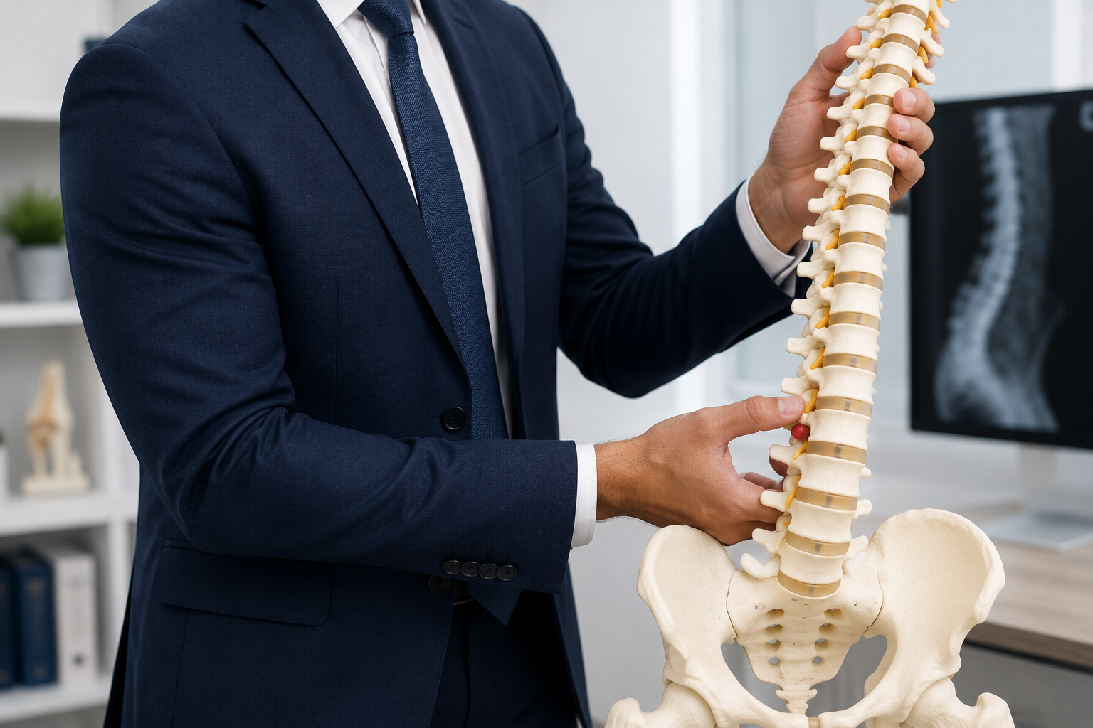
★ Featured guide · 8 min read
Understanding Your Spine — Patient Leaflet
Full illustrated guide to spinal anatomy — the bones, discs, joints, ligaments, muscles and nerves, written in plain English for patients.
For information only — not medical advice. The content on this page is patient education to help you understand a condition or treatment. It is not a substitute for personalised assessment by a qualified clinician. Always consult your doctor or specialist about your own symptoms, diagnosis, and treatment decisions.
A guide to the anatomy
Your spine is one of the most remarkable structures in the human body. It holds you upright, lets you bend, twist and reach, absorbs the shocks of everyday movement, and protects the delicate nerves that carry signals between your brain and the rest of you — all at the same time. We tend to picture the spine as a single bone running down the back, but it is really a column of many different parts, each with its own job, working together as one. Understanding how those parts fit together makes it far easier to make sense of back and neck problems, and of the treatments that help them.
This guide walks through the anatomy of the spine, one structure at a time.
The spine as a whole
Seen from the front or back, the spine is a tall column of small bones stacked one on top of another, running from the base of the skull to the pelvis. Seen from the side, it is not straight but gently S-shaped, with curves that sweep inward at the neck and lower back and outward through the chest and pelvis.
Those curves are not a flaw. They behave like the curve of a spring, spreading load evenly and softening the jolts of walking and running. A perfectly straight column would transmit every bump straight up to the head; a gently curved one absorbs and distributes it. The whole structure has to manage a difficult balancing act — it must be strong enough to carry the weight of your head and trunk, yet flexible enough to let you move freely. The way the spine is built lets it do both.
The regions of the spine
The spine is divided into five regions, working from top to bottom: the cervical spine (neck) — seven vertebrae that support the head and allow it to nod, tilt and turn, the smallest and most mobile of the spinal bones; the thoracic spine (upper and mid back) — twelve vertebrae, each connected to a pair of ribs, built more for stability than movement and forming the back wall of the chest; the lumbar spine (lower back) — five large vertebrae that carry the most weight and take much of the strain of bending and lifting, where back pain most commonly arises; the sacrum — five vertebrae that fuse together in adulthood to form a single solid bone, wedged between the two halves of the pelvis; and the coccyx (tailbone) — the small bones at the very base of the spine, the remnant of a tail.
Each region has a slightly different shape and purpose, but the basic design of the building blocks is shared throughout.
Vertebrae: the building blocks
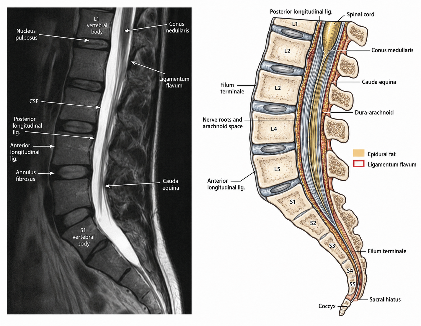
The individual bones of the spine are called vertebrae. Each one has two main parts. At the front is the vertebral body — a solid, drum-shaped block that bears the weight, rather like one reel in a stack of cotton reels. At the back is a ring of bone, made up of the pedicles and lamina, that arches behind the body. When the vertebrae are stacked, these rings line up to form a continuous bony tunnel called the spinal canal, through which the spinal cord runs.
Projecting from the back of each ring are several bony points known as processes. The spinous process is the knobble you can feel running down the middle of your back; the transverse processes point out to each side; and the articular processes link up with the vertebrae above and below. These act as handles and levers — the anchor points that muscles and ligaments pull on to move and steady the spine.
In short, every vertebra is both a load-bearing pillar at the front and a protective, movable arch at the back.
Intervertebral discs: the shock absorbers
Between most of the vertebrae sits a tough, springy cushion called an intervertebral disc. The discs are the spine's shock absorbers, spreading the forces generated by walking, running, lifting and jumping so they don't hammer the bones directly.
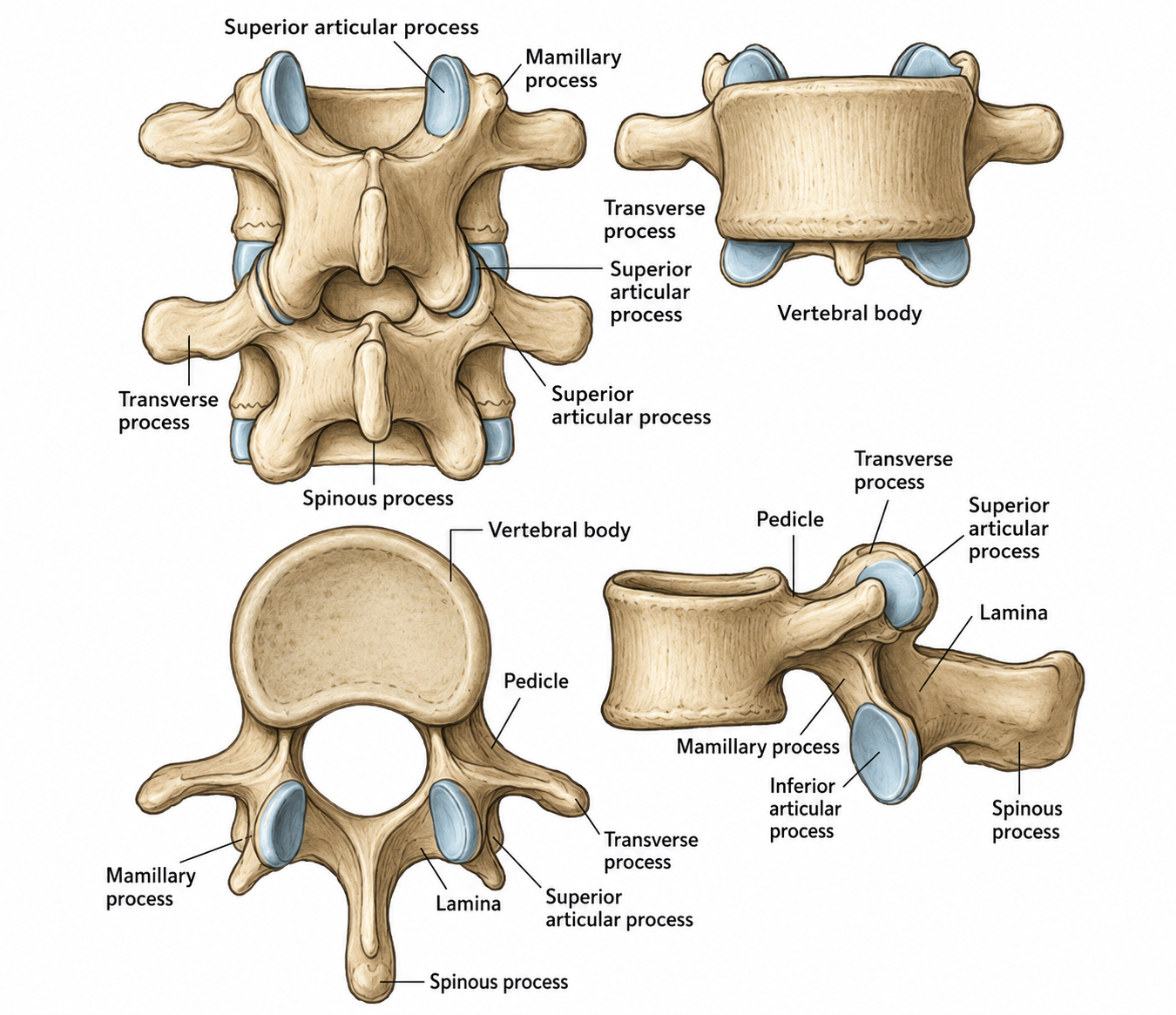
Each disc has two parts that work together. The annulus fibrosus is the strong outer ring, built from many layers of tough fibres laid in criss-crossing rings — a little like the plies of a car tyre, or the rings of an onion. It holds the disc together and contains the centre. The nucleus pulposus is the soft, gel-like core. In younger people it is rich in water and behaves rather like a water balloon, squashing and reshaping under pressure to spread the load. A useful way to picture the whole disc is as a jam doughnut: a firm, layered outer ring wrapped around a soft jelly centre.
Discs are unusual in the body because, deep inside, they have almost no blood supply, no nerve supply and no drainage system. In fact, the disc is the largest structure in the body that survives without its own direct blood supply, feeding slowly by nutrients soaking in from its edges. This is simply part of how a healthy disc is built — but it is also why discs behave differently from most other tissues, drying out and stiffening gradually over the years as a normal part of ageing.
Facet joints: the guides
At the back of each vertebra are small paired joints called facet joints, which link each vertebra to its neighbours above and below. They guide and steer the spine's movement — controlling how far you can bend forward, arch backward and twist — while also acting as stops that prevent one vertebra from sliding too far on the next.
Like the knee or the knuckles, facet joints have smooth cartilage surfaces and a little lubricating fluid, which lets them glide easily. A helpful image is the guide rails on a sliding drawer, or the hinges on a door: they allow movement in the directions the spine is designed for, while blocking movement it shouldn't make.
Ligaments: the stays
Ligaments are strong, slightly elastic bands of tissue that tie the bones of the spine together. They run along the front, the back and the inside of the spinal column, holding everything in good alignment and stopping the spine from moving beyond its safe limits.
You can think of them as the stays and guy-ropes that hold up a tall, flexible mast — taut straps that keep the column upright and steady, while still allowing it to flex. Working as a team, the ligaments quietly provide a great deal of the spine's stability.
Muscles: the active support
Bones and ligaments give the spine its passive framework, but the muscles around it provide active, moment-to-moment support. The deep muscles of the trunk and back help maintain posture, power and control movement, protect the spine from sudden strain, and keep you balanced and coordinated.
Many of these deep muscles wrap around the spine like a natural, built-in corset, constantly tensing and relaxing to brace the column — much as a crew keeps adjusting the rigging on a boat to hold it steady. Strong, well-conditioned muscles take a great deal of load off the discs and joints, which is one reason the muscular system is such an important part of spinal anatomy.
The spinal cord
Running down through the protective bony canal is the spinal cord, one of the most important structures in the entire body. It is the main communication pathway between the brain and everything below, carrying messages up and down so that you can move, feel sensation and coordinate your body.
In adults the cord itself does not run the full length of the spine. It ends at around the level of the first lumbar vertebra, tapering to a point called the conus medullaris. Below that point, the nerves continue downward as a loose bundle of roots known as the cauda equina — Latin for "horse's tail," which is exactly what it resembles. You can picture the cord as the body's main cable, like the trunk line carrying signals between a control centre and everywhere it serves, with the bony canal acting as its armoured conduit.
Spinal nerves and nerve roots
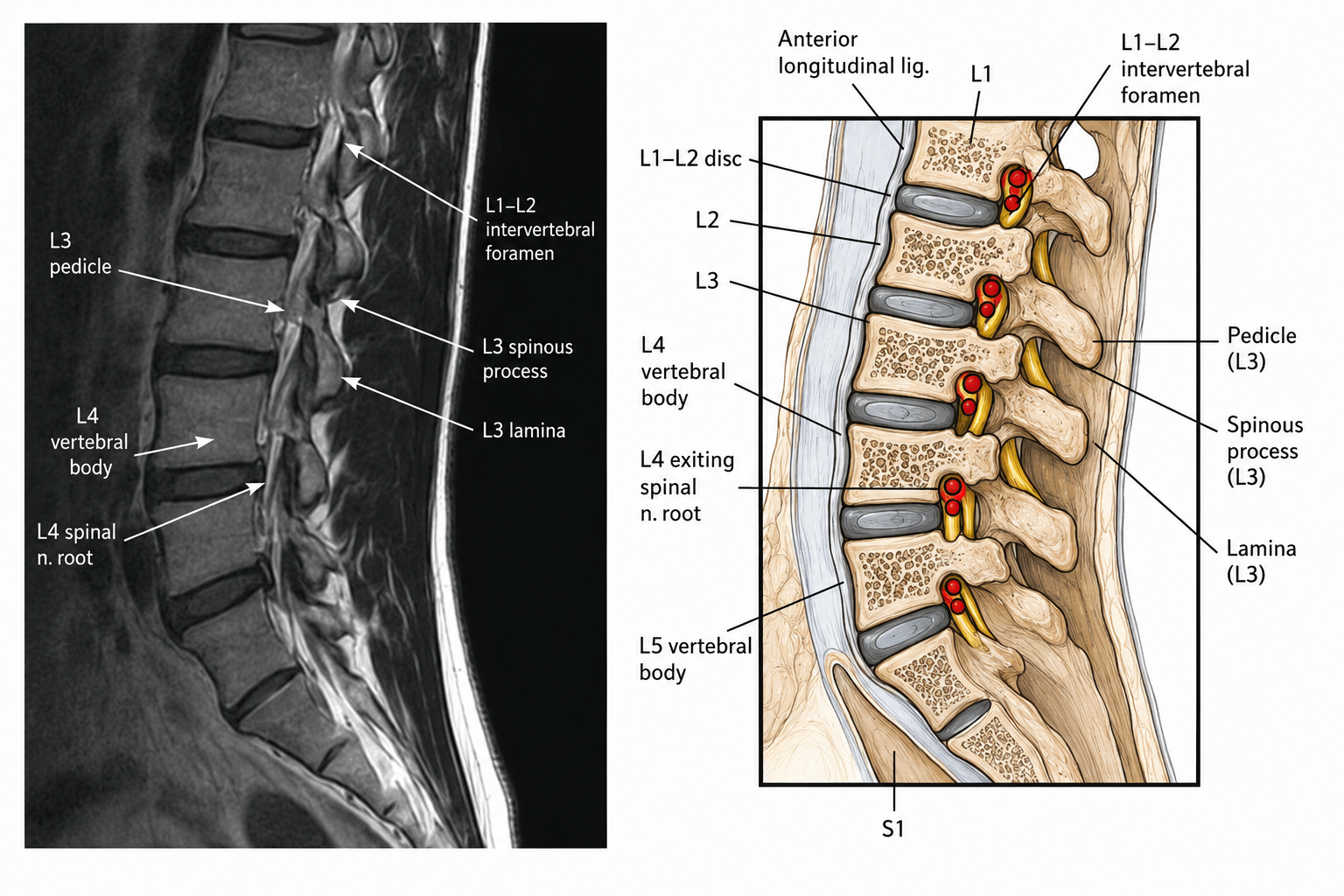
At every level of the spine, pairs of nerves branch off the spinal cord and leave the spinal canal through small openings between the vertebrae called intervertebral foramina. These spinal nerves then travel out to the body, and each is responsible for muscle strength, sensation, reflexes and coordination in a particular area.
The foramina act rather like the exit doorways, or the off-ramps from a motorway, where each nerve leaves the spine to reach its destination. Because each nerve serves a specific region, the pattern of where symptoms are felt can tell a great deal about which level of the spine a nerve is travelling from — useful information when working out the source of a problem.
What bone is made of
Finally, it is worth understanding the material the vertebrae themselves are built from, because bone is not the dry, lifeless substance it is often imagined to be. Bone is living tissue, constantly being renewed by two teams of cells: osteoblasts, which build new bone, and osteoclasts, which clear away old bone. Throughout life these two crews keep the skeleton repaired and renovated.
Bone gets its strength from two ingredients working together. A flexible protein scaffold, made mainly of collagen, gives bone the ability to bend a little without snapping, while a hard mineral — largely calcium — is packed onto that scaffold to give stiffness and resistance to crushing. The combination behaves much like reinforced concrete, where steel reinforcing bars provide flexibility and the surrounding concrete provides hardness; together they are far stronger than either alone.
Inside a vertebra, a thin, dense outer shell of cortical bone wraps around an inner lattice of fine bony struts called trabecular bone, arranged like a honeycomb or internal scaffolding. This honeycomb is astonishingly strong for its weight, and its struts are cleverly aligned along the very directions in which force passes through the bone. It is this hidden internal structure that allows your vertebrae to carry the load of the body day after day.
How it all works together
Each of these structures is remarkable in its own right, but the real elegance of the spine lies in how they combine. The vertebrae provide the pillars, the discs cushion them, the facet joints steer the movement, the ligaments hold the alignment, and the muscles brace and power the whole column — all while the bony canal and the foramina protect the spinal cord and the nerves running through and out of it.
It is a structure built to be both strong and supple, and to keep working reliably over a lifetime of movement. Understanding how your spine is put together is the first step to understanding how to look after it, and what can be done when something isn't working as it should.
Patient downloads
Take this away
Print-friendly guides and forms you can save, share with your GP, or bring to your consultation. All documents are written for patients in plain English.
References & further reading
- —NICE — Low Back Pain and Sciatica in Over 16s (NG59)
- —NHS — Spinal Conditions
- —British Association of Spine Surgeons (BASS)
This information is for general patient education and is not a substitute for an individual consultation. Always seek personalised advice from a qualified spinal specialist.
Found this helpful? Share it
Discuss this with Mr Madi
Personalised, consultant-led advice
Every patient is different. The information on this page is a starting point — Mr Madi will tailor any plan to your specific imaging, symptoms and goals.